
Fichièr:Ebola virus (2).jpg

Fichièr d'origina (2 365 × 1 843 pixèl, talha del fichièr: 572 Ko, tipe MIME: image/jpeg)
| Aqueste fichièr proven de Wikimedia Commons?. Las informacions que lo concernisson son afichadas çaijós (procedura). |
.jpg?uselang=oc)
Descripcion
| Descripcion |
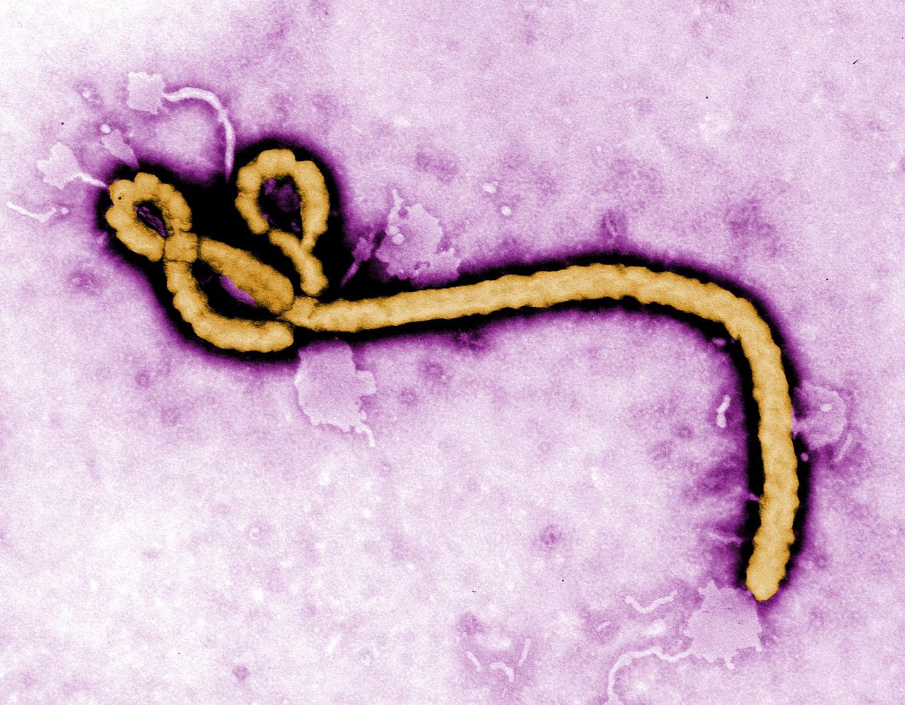
Created by CDC microbiologist Frederick A. Murphy, this colorized transmission electron micrograph (TEM) revealed some of the ultrastructural morphology displayed by an Ebola virus virion. See PHIL 1181 for a black and white version of this image. What is Ebola hemorrhagic fever (Ebola HF)? Ebola hemorrhagic fever (Ebola HF) is a severe, often-fatal disease in humans and nonhuman primates (monkeys, gorillas, and chimpanzees) that has appeared sporadically since its initial recognition in 1976. The disease is caused by infection with Ebola virus, named after a river in the Democratic Republic of the Congo (formerly Zaire) in Africa, where it was first recognized. The virus is one of two members of a family of RNA viruses called the Filoviridae. There are four identified subtypes of Ebola virus. Three of the four have caused disease in humans: Ebola-Zaire, Ebola-Sudan, and Ebola-Ivory Coast. The fourth, Ebola-Reston, has caused disease in nonhuman primates, but not in humans. For more information on Ebola and what CDC is doing, please visit: www.cdc.gov/vhf/ebola/ |
| Data | |
| Font | Ebola virus |
| Autor | CDC Global |
Publicat jos licéncia(s)
- Sètz liure :
- de partejar – de copiar, distribuir e transmetre aquesta òbra
- d'adaptar – d'adaptar aquesta òbra
- Jos las condicions seguentas :
- atribucion – Vos cal atribuir aquesta òbra amb lo biais especificat per l'autor o lo concedent (mas pas dins un sens que suggerís que vos apròvan o qu'apròvan l'utilizacion d'aquesta òbra).
| Cette image a été originellement postée sur Flickr par CDC Global Health à l'adresse https://www.flickr.com/photos/89075068@N07/14723720857. Elle a été passée en revue le 10 octobre de 2014 par le robot FlickreviewR, qui a confirmé qu'elle se trouvait sous licence cc-by-2.0. |
Istoric del fichièr
Clicar sus una data e una ora per veire lo fichièr tal coma èra a aqueste moment
| Data e ora | Miniatura | Dimensions | Utilizaire | Comentari | |
|---|---|---|---|---|---|
| actual | 9 octobre de 2014 a 22.03 | | 2 365×1 843 (572 Ko) | Discasto | Transferred from Flickr via Flickr2commons |
Paginas que contenon lo fichièr
La pagina çaijós compòrta aqueste imatge :
Usatge global del fichièr
Los autres wikis seguents utilizan aqueste imatge :
- Utilizacion sus ca.wikinews.org
- Utilizacion sus eu.wikipedia.org
- Utilizacion sus fr.wikipedia.org
- Virus Ebola
- Vaccin contre le virus Ebola
- Liste d'épidémies liées au virus Ebola
- 3-Déazaneplanocine A
- Épidémie de maladie à virus Ebola en Afrique de l'Ouest
- Maladie à virus Ebola
- Favipiravir
- Brincidofovir
- FGI-106
- BCX4430
- LJ-001
- FGI-104
- FGI-103
- TKM-Ebola
- JK-05
- CAd3-ZEBOV
- VSV-EBOV
- Ameyo Adadevoh
- Première épidémie de virus Ebola de 2018 en république démocratique du Congo
- Épidémie de maladie à virus Ebola au Kivu
- Modèle:Palette Ebola
- Massacre de Womey
- Portrait sur EPI
- Projet:Aide et accueil/Twitter/Tweets/archives/août 2020
- Utilizacion sus he.wikipedia.org
- Utilizacion sus mg.wikipedia.org
- Utilizacion sus pl.wiktionary.org
- Utilizacion sus so.wikipedia.org
- Utilizacion sus species.wikimedia.org
- Utilizacion sus uk.wikipedia.org
.jpg)